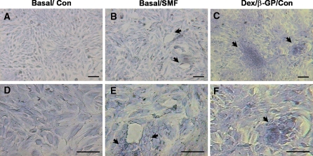
Fig. 3.
The cell morphology and the expression of extracellular matrix in cells exposed to SMF (290 mT, average flux density) for 7 days. The metachromatic staining of the extracellular matrix with toluidine blue was observed with different magnification of images for the unexposed (a,d) and SMF-exposed group (b,e) in basal culture medium. The cells cultivated with osteogenic medium served as the positive control (Dex/β-GP/Con) to compare the SMF-mediated effect (c,f). The arrows indicated the special morphology (b,c) and the obvious matrix condensation (e,f) around the circle of cell arrangement to compare with non-exposed cells with basal culture medium. The scale bar represents 50 μm. (Con) control, unexposed group; (SMF) static magnetic field; (Basal) basal medium; (Dex/β-GP) dexamethasone/β-glycerophosphate

