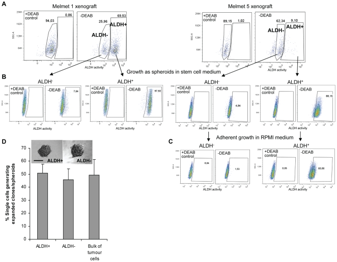
Figure 2. In vitro comparison of clonogenic potential of ALDH+ and ALDH− cells isolated from melanoma xenografts.
(A) ALDH+ and ALDH− subpopulations identified in Melmet 1 and Melmet 5 xenografts. (B) Evaluation of the ALDH phenotype in the daughter spheroids formed during culturing of sorted ALDH+ and ALDH− cells in hESCM as described in (D). (C) Aldefluor analysis of the spheroid-derived cells subsequently cultured adherently in RPMI for 2 weeks (representative dot-plots only for Melmet 5). (D) Sorted ALDH+ and ALDH− cells were seeded one cell/well and cultured in hESCM to allow formation of single-cell-derived clones, spheroids showed in the insert (bar, 100 µm). Efficiency of spheroid formation from unsorted bulk melanoma cells is presented for comparison. Data represents mean ± SEM (n = 7). The formed spheroids from each group were collected, disintegrated into single cells that were further cultured in hESCM for formation of daughter spheroids. The latter were reanalyzed by the Aldefluor assay as shown in (B) or cultured further in RPMI as shown in (C).

