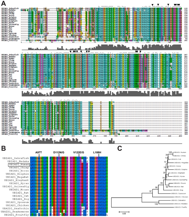
Figure 2. Highly conserved UBIAD1 residues are mutated in SCD.
(A) Locations of 17 amino acids mutated in SCD patients are indicated by arrows. Taller bars in the graph below the alignment indicate greater conservation. (B) Regions of alignment encompassing human SCD mutations: A97, D112, V122, L188, are shown. The position of the human protein in the alignment is indicated on the left (box). (C) Evolutionary relationships based upon UBIAD1 homology.

