Figure 3.
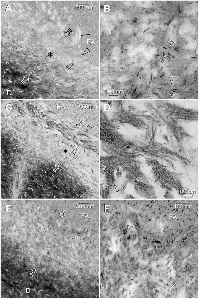
TEM images of control (A–B) and experimental (C–F) dentin specimens bonded with Adper Scotchbond SE. FA: filled adhesive; D: dentin; T: dentinal tubule; Between open arrows: the original 2Δm thick, partially demineralized interdiffusion zone. A. A control specimen that was retrieved after immersion in the Ca2+ and PO43−-containing simulated body fluid (SBF) for the entire experimental period. There was an increase in the thickness and density of the partially-demineralized region within the interdiffusion zone, probably due to epitaxial growth over remnant seed crystallites (see Fig. 1C). However, in the absence of biomimetic analogs, there was no remineralization of the surface, completely-demineralized part of the interdiffusion zone (between open arrowheads). The surface collagen fibrils (arrow) were electron lucent (i.e. devoid of minerals). B. A high magnification view of the location marked by the asterisk in Fig. 3A. Remineralization was predominantly extrafibrillar along the surface of the collagen fibrils (between open arrowheads). Intrafibrillar remineralization could not be recognized, probably due to depletion of remnant intrafibrillar seed crystallites from this location. C. An experimental specimen that exhibited features of an early stage of biomimetic remineralization. Unlike the control specimen (Fig. 3A), the surface part of the interdiffusion zone (between open arrowheads) was remineralized when biomimetic analogs are included in the SBF. The mode of remineralization was intrafibrillar in nature and consisted of the electron dense braided mineral phase that at this stage appeared completely different from the mineral phase present in the underlying interdiffusion zone (asterisk). D. A high magnification view of the electron dense braided mineral phase in Fig. 3C. In some collagen fibrils, the continuous electron dense phase had been transformed into discrete nanocrystals that followed the braided appearance of the microfibrillar strands (open arrowhead). E. An experimental specimen that exhibited features of a more mature stage of biomimetic remineralization. Minerals were present within the entire interdiffusion zone. Remnant braided mineral phases (arrows) could still be identified along the superficial part of the interdiffusion zone. F. A high magnification view of the superficial part of the interdiffusion zone showing the presence of mineral platelets (arrows) within a region that was completely-demineralized in the baseline (Fig. 1C) and control specimens (Fig. 3A). Remnants of the nanocrystals (open arrowheads) could be identified within this region.

