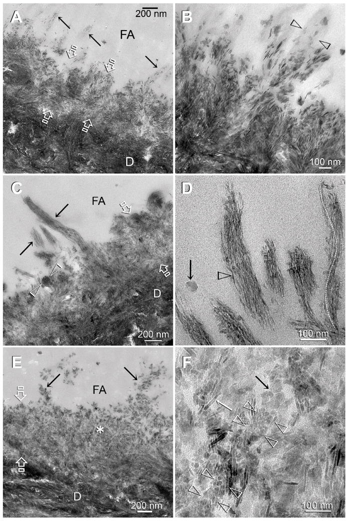
Figure 4.
TEM images of control (A–B) and experimental (C–F) dentin specimens bonded with Adper Easy Bond. FA: filled adhesive; D: dentin; T: dentinal tubule; Between open arrows: the original 0.5 Δm thick, partially demineralized interdiffusion zone. A. In the absence of biomimetic analogs, remineralization of the interdiffusion zone probably occurred via epitaxial growth over remnant intrafibrillar and extrafibrillar seed crystallites. Some of the tufted collagen fibrils along the surface of the interdiffusion zone (arrows) also appeared to have remineralized. B. A high magnification view of those surface collagen fibrils in the control specimen showing that remineralization was extrafibrillar in nature, along the some parts surface of the collagen fibril (between open arrowheads), while other parts of the same fibril were not remineralized. C. An experimental specimen that exhibited features of an early stage of biomimetic remineralization. Collagen fibrils within the entire interdiffusion zone contained the electron dense braided mineral phase, masking the existing apatite platelets from the original partially demineralized part of the interdiffusion zone. The electron-dense braided mineral phase was more easily observed from the tufted surface collagen fibrils (arrows). A collagen fibril that was sectioned transversely (between open arrowheads) clearly indicated that the entire fibril was filled with intrafibrillar minerals. D. A high magnification view of the remineralized, tufted collagen fibrils similar to those shown in Fig. 4C indicated that the electron-dense braided mineral phase remained continuous at this stage (open arrowhead). This electron-dense mineral phase followed the helical arrangement of the microfibrils (white dotted line). The electron-dense, amorphous structure (arrow) adjacent to the remineralized fibrils could represent a fluidic amorphous calcium phosphate nanoprecursor droplet (see Fig. 2B). E. An experimental specimen that exhibited features of a more mature stage of biomimetic remineralization. At this magnification, the bulk of the interdiffusion zone and the tufted surface collagen fibrils (arrows) were filled with mineral platelets. The electron-dense braided mineral phase could no longer be observed. F. A high magnification view taken from the location marked by the asterisk in Fig. 4E. Interpretation of the micrograph was complicated by the superimposition of remineralized minerals over existing minerals. A longitudinal section through a collagen fibril (between arrowheads) showed that it was filled with miniature mineral platelets that were much smaller than some of the adjacent platelets (arrows). Those miniature mineral platelets probably represented the transformed platelet phase from the continuous electron-dense braided mineral phase (see Fig. 3D).

