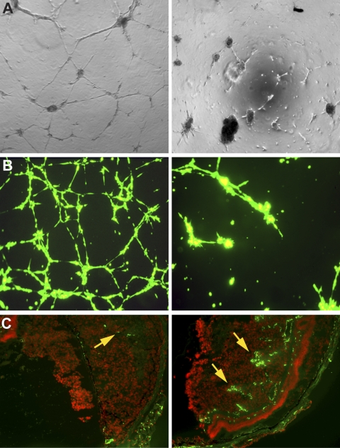
Figure 2.
Bevacizumab decreased angiogenesis in vitro and in vivo. Bevacizumab decreased tube formation: IgG1-treated HUVECs (A, left) and Mel290-GFP co-cultured with HUVECs (B, left) exhibited meshlike patterns of tube formation (grade 5). Corresponding cells treated with bevacizumab showed a migration/alignment pattern (grade 1) in HUVECs (A, right) and sprouting new capillary tubes (grade 3) in the melanoma-GFP co-cultured cells (B, right). Bevacizumab decreased the vascular density in our mouse model of ocular melanoma (C). CD31+ vascular channels constitute 1% of the area of the tumor in mice treated with bevacizumab 250 μg/mL (C, left, arrow), compared with 26% of the area of the tumor in control mice treated with PBS (C, right, arrows).

