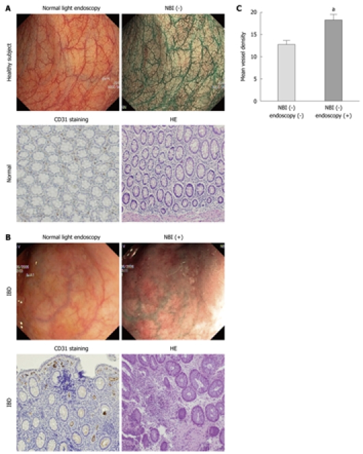
Figure 1.

Colonic mucosa of healthy individuals and uninflamed but narrow-band imaging (NBI)+ regions from patients with inflammatory bowel disease (IBD), visualized using white light colonoscopy and NBI endoscopy. The microvasculature of histologically normal control and IBD uninflamed colonic mucosa was immunohistochemically stained for CD31 and von Willebrand/factor VIII. There was increased vascularization in uninflamed IBD mucosa that presented an aberrant NBI+ pattern, in the NBI+ areas compared with controls. A: Healthy subjects. The vascular pattern was normal on both conventional colonoscopy and NBI, which was confirmed by immunohistochemical staining; B: Areas of the IBD mucosa that were not inflamed on conventional colonoscopy and showed an aberrant NBI+ pattern. Immunohistochemical staining confirmed an increased vascularization in these areas compared with NBI- areas; C: Computerized morphometric analysis of the microvasculature in control and IBD uninflamed mucosa that was NBI+. After immunohistochemical staining, sections were analyzed for the total number of vessels/field (microvascular density). aA statistically significant difference was found between sections from areas of uninflamed IBD colonic mucosa that were NBI+ and from controls.
