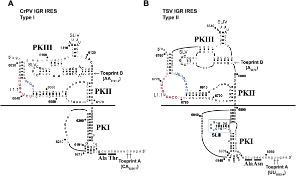
FIGURE 1.
The secondary structure of the (A) Type I CrPV IGR IRES and the (B) Type II TSV IGR IRES. Conserved nucleotide positions are shown in uppercase and nonconserved nucleotides are in lowercase. Numbering refers to the nucleotide position within the respective viral genome. Helical regions are indicated by a black dash between nucleotides. Underlined nucleotides represent the first two amino acid residues in the viral capsid protein. Properly positioned 40S and 80S ribosomes on the IGR IRES produces a toeprint, denoted Toeprint A, shown by the arrows. The point at which the two classes of IGR IRES were swapped is indicated by a black line. The ΔSLIII deletion within the Type II TSV IGR IRES is marked by a blue box. L1.1A and L1.1B are indicated by red and blue letters, respectively.

