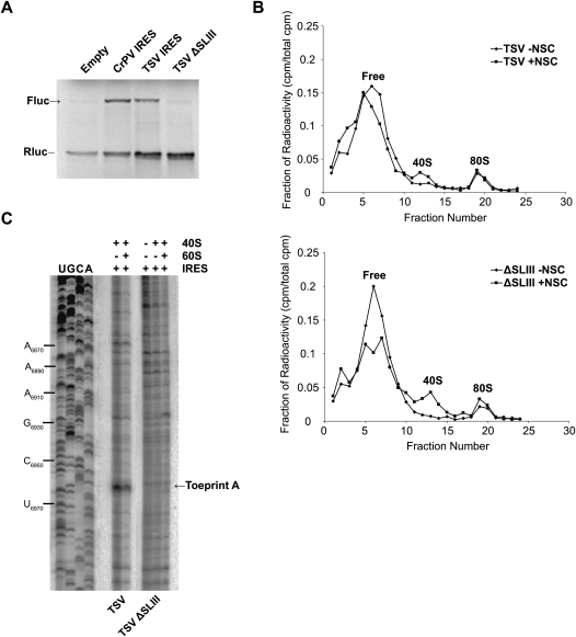
FIGURE 7.
Characterization of SLIII within the TSV IGR IRES. (A) Dicistronic RNAs containing wild-type or ΔSLIII TSV IGR IRESs were incubated in RRL at 30°C for 60 min in the presence of [35S]methionine. The first cistron, encoding Renilla luciferase (Rluc), measures scanning-mediated translation, and the second cistron, firefly luciferase (Fluc), measures IGR IRES-mediated translation. Shown are radiolabeled firefly (Fluc) and Renilla (Rluc) luciferase protein products detected by autoradiography and quantitated by PhosphorImager analysis. (B) 80S assembly on TSV and ΔSLIII IGR IRESs was assessed in RRL by sucrose gradient analysis. Radiolabeled wild-type or ΔSLIII IGR IRES (100 nM) was incubated in RRL with 0.1 mg/mL cycloheximide in the presence (+NSC) or absence (−NSC) of 25 μM NSC119889 for 15 min at room temperature. Reactions were loaded on a 10%–30% sucrose gradient and shown are the percent of total radioactive counts in each fraction. The top and bottom of the gradient is represented from left to right, respectively. Fractions containing free IRES, 40S, and 80S ribosomes are indicated. (C) Toeprint analysis of assembled 40S and 80S ribosomes on TSV and ΔSLIII IGR IRESs. 40S alone or 40S and 60S subunits (100 nM) were incubated with dicistronic RNAs containing wild-type or mutant IGR IRES and analyzed by primer extension analysis using oligo PrEJ69. Reaction products were separated in denaturing polyacrylamide gels. The gels were dried and exposed by autoradiography.

