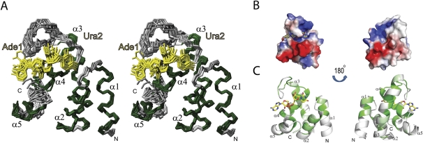
FIGURE 4.
Solution structure of the human PHAX-RBD bound to AUCG RNA. (A) Stereo view of the 10 lowest energy structures of PHAX-RBD bound to AUCG. Residues 224–311of PHAX-RBD and the first two bases of the RNA, Ade1 and Ura2, are shown. Helical secondary structure elements are colored in green and loops in gray. RNA bases are colored in yellow. (B) Electrostatic surface representation of PHAX-RBD. (C) Sequence conservation of the PHAX-RBD in metazoan species plotted onto a surface representation of the structure. Colors from white to green correspond to increasing conservation.

