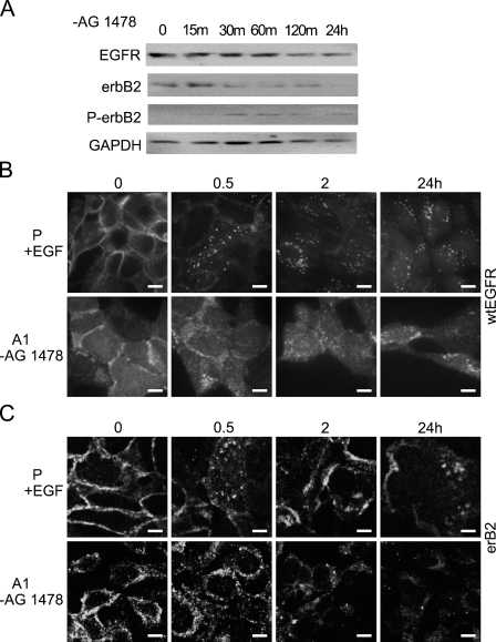
Figure 4.
Examination of protein levels and cellular localization of wtEGFR and erbB2 as a result of acute activation of EGFRvIII. vIIIA1 (A1) cells were treated with (+)AG1478 for 7 days, and then EGFRvIII was acutely activated by removing (−)AG1478 for the indicated times. (A) Immunoblot analysis of wtEGFR, erbB2, and phospho-erbB2 (P-erbB2) in cells. GAPDH was detected as an internal loading control. Confocal images show localization of (B) wtEGFR and (C) erbB2 in EGFRvIII-expressing cells after acute activation of EGFRvIII for the indicated times, compared with activation of wtEGFR in the parental (P) cell line for the indicated times. P, OVCA 433 parental cells; A1, vIIIA1 cells; m, min; h, hr. Bar = 10 μm.

