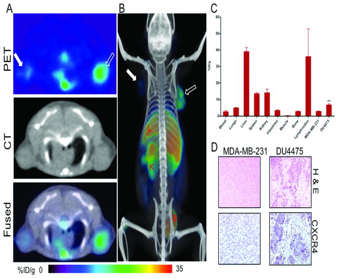
Figure 3. CXCR4 imaging in orthotopic breast tumor xenografts with [64Cu]AMD3100.
NOD/SCID mice harboring MDA-MB-231 and DU4475 orthotopic breast tumor xenografts in the upper thoracic mammary fat pad received ~11 MBq of [64Cu]AMD3100 via tail vein injection and whole body images were acquired at 90 min post-injection. A, transaxial PET, CT and fused sections of both the tumors; B, volume rendered whole body image showing clear accumulation of radioactivity in DU4475 tumor; C, biodistribution analysis of selected tissues from mice injected with 740 kBq of [64Cu]AMD3100 and sacrificed at 90 min post-injection. All radioactivity values were converted into percentage of injected dose per gram of tissue (%ID/g). Biodistribution data are means ± SEM of four to five animals; D, representative microscopy images of 10 μm-thick hematoxylin and eosin and CXCR4 stained sections obtained at ×10 magnification from both tumors. Significance is indicated by asterisks (*) and the comparative reference is MDA-MB-231 tumor uptake. ***P < 0.001. Solid arrow, MDA-MB-231 tumor; open arrow, DU4475 tumor; L, liver; K, kidney; B, bladder.

