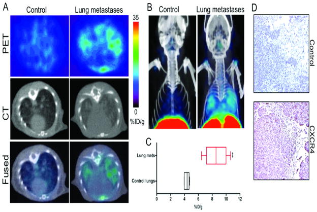
Figure 4. CXCR4 imaging of MDA-MB-231 derived lung metastases with [64Cu]AMD3100.
NOD/SCID mice that received either 2×10⁶ MDA-MB-231 cells or HBSS were received ~11 MBq of [64Cu]AMD3100 at 35 days after inoculation. Whole body images were acquired at 90 min after injection. A, transaxial PET, CT and fused sections of lung metastasis and control mice; B, volume rendered whole body image showing clear accumulation of radioactivity in the lung metastases. Top slices of the volume rendered images were cut for clear visualization of lung uptake; C, box-and-whisker plot of the biodistribution analysis of lungs from mice injected with 740 kBq of [64Cu]AMD3100 at 90 min post-injection. All radioactivity values were converted into percentage of injected dose per gram of tissue (%ID/g) and are means ± SEM of four to five animals; D, representative microscopy images of 10 μm-thick CXCR4 and control antibody stained sections of the lung metastases obtained at ×10 magnification. Significance is indicated by asterisks (*) and the comparative reference is the lungs from mice injected with HBSS. ***P < 0.001.

