
Fig. 5.
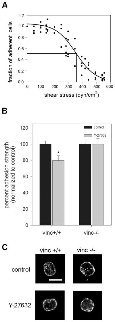
Loss of vinculin abolishes differences in adhesion strength due to contractility independent of bound integrin levels. A, Characteristic detachment profile for vinculin+/+ and vinculin−/− MEFs showing the fraction of adherent cells (f) vs. applied shear stress (τ). These cells exhibit the same detachment profiles as the NIH3T3 cells. B, vinculin +/+ and vinculin −/− MEFs were cultured for 16 hours on micropatterned surfaces and treated with Y-27632 (50μM) for 30 minutes prior to spinning. The no-treatment group received fresh media without inhibitor. Addition of inhibitor resulted in a 20% decrease in adhesion strength for the vinculin +/+ cells. No differences in adhesion strength were detected for the vinculin −/− cells. *p<0.03 relative to no treatment vinculin +/+ MEFs. C, Immunoflourescence staining for α5 integrin subunit following cross-linking/extraction showing localization of integrins mostly at the periphery of the substrate. Treatment with Y-27632 (50μM) for 30 minutes did not significantly alter integrin binding.