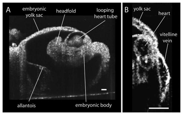
Fig. 1.

Live structural imaging of 8.5 day mouse embryo with SS-OCT. A, Typical 3D reconstruction of the whole embryo with the yolk sac. B, SS-OCT image depicting a cross section of a heart and a fragment of vitelline vein with individual circulating blood cells. The scale bars correspond to 100 μm.
