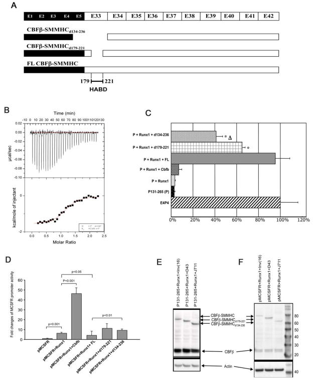
Figure 1. The type I CBFβ-SMMHC fusion variant is very inefficient in binding and repressing RUNX1.
(A) Diagrammatic representation of CBFβ-SMMHC fusion variants. CBFβ-SMMHCd134-236, generated by type I fusion, misses the HABD as well as CBFβ residues encoded by exon 5. CBFβ-SMMHCd179-221 misses only the HABD and has been described before (Lukasik et al., 2002).
(B) ITC measurement of interactions between 200 μM CBFβ-SMMHCd134-236 and 7.5 μl injections of 15 μM RUNX1 Runt domain. The top panel shows the raw data while the bottom panel is a plot of the binding corrected for the dilution enthalpy (average dilution enthalpy = 232 cal mol−1). Data were fit to a one-site binding model. The results of a fit to one titration are shown in the box at the lower right corner. The average Kd of two independent experiments is 709 (± 47) nM.
(C) CD4 reporter assay. E4P4: CD4 enhancer and promoter; P131-265: core sequence of CD4 repressor; FL: CBFβ-SMMHC; d179-221: CBFβ-SMMHCd179-221; d134-236: CBFβ-SMMHCd134-236. Δ: statistically significant difference (P<0.05) between the top two conditions, which are P131-265 + Runx1 with either d179-221 or d134-236. *: statistically significant differences (P<0.05) between the top two conditions and the third one, which is P131-265 + Runx1 + FL.
(D) MCSFR reporter assay. pMCSFR: the luciferase reporter driven by human MCSFR promoter.
(E and F) Western blot analysis showing the expression of the transfected constructs in the reporter assays (C and D, respectively). The expression of CBFβ and full-length and variant CBFβ-SMMHC constructs was detected with a mouse monoclonal antibody (β141.2) specific for CBFβ.
The error bars in (C) and (D) represent one standard deviation.

