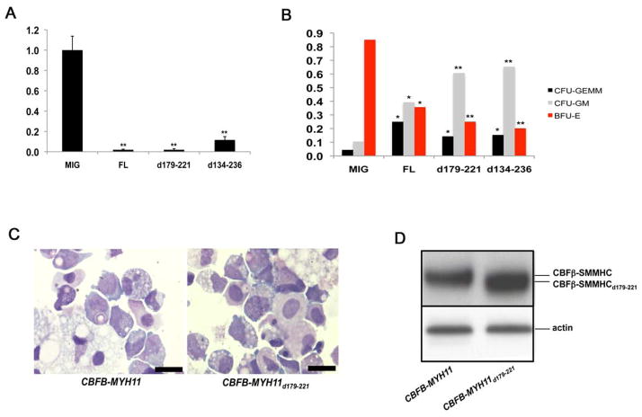
Figure 5. Growth and differentiation defects of human CD34+ cells transduced with CBFB-MYH11 variants.
(A) and (B) In vitro differentiation of human CD34+ cells. Human CD34+ cells were transduced with empty retroviral vector (MIG), and vectors expressing CBFB-MYH11 (FL), CBFB-MYH11d179-221 (d179-221), or CBFB-MYH11d134-236 (d134-236). The transduced cells were sorted by FACS and two thousand GFP+ cells were plated in serum-free methylcellulose cultures. (A) Total colony numbers scored after 14 days. The total colony number from MIG-transduced cells was set at 1, and the total colony numbers from the other two transduced cell populations were calculated relative to that of MIG. The data shown are averages of three independent experiments. (B) Frequencies of BFU-E, CFU-GM, and CFU-GEMM colonies from transduced human CD34+ cells. The data shown are averages of two independent experiments. *: p<0.01 between clones transduced with MIG and those with CBFB-MYH11 constructs; **: p<0.001 between clones transduced with MIG and those with CBFB-MYH11 constructs. The error bars represent one standard deviation.
(C) Cell morphology from long-term cultures of human CD34+ cells transduced with CBFB-MYH11 or CBFB-MYH11d179-221. Shown are cytospin preparations of non-adherent cells at week 10, stained with Wright-Giemsa stain. Scale bars represent 10 μm.
(D) Western blot analysis with protein samples from transduced human CD34+ cells and the CBFβ-specific antibody (β141.2).

