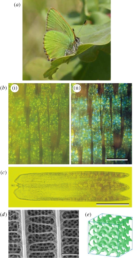
Figure 1.
(a) C. rubi in the resting position on an oak leaf, showing the green-coloured ventral face of its wings (photography Bea Koetsier). The green colour ensures excellent camouflage in its natural habitat. (b) Optical micrograph of a small region of ventral wing scales taken in reflection. b(i) Illumination with linearly polarized white light while the image was captured through a parallel linear analyser; b(ii) same as in b(i) but the image was captured through a crossed linear analyser. Green, blue and yellow spots can be identified. Scale bar, 100 µm. (c) Optical micrograph of a single ventral wing scale taken in transmission. Several domains are observed across the distal part of the scale. The dark longitudinal lines are the ridges. Scale bar, 50 µm. (d) Scanning electron micrograph of part of a scale. Scale bar, 1 µm. Below the network of ridges and crossribs, a three-dimensional cuticular structure is seen. (e) Gyroid structure modelling the cuticular structure (16 cubic unit cells).

