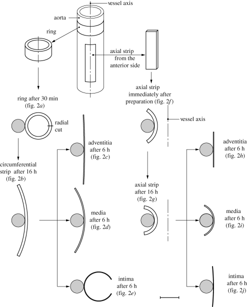
Figure 1.
Schematic of the procedure for specimen preparation showing: ring and axial strip specimens from the aorta, after 30 min of equilibration and immediately after preparation; circumferential and axial strips after 16 h of equilibrium; circumferential and axial strips from the separated intima, media and adventitia after 6 h of equilibrium. Each specimen was glued to a plastic tube. The various configurations are shown to the correct scale. Scale bar: 10 mm. Adapted from fig. 2 of Holzapfel et al. (2007) with kind permission of Springer Science and Business Media.

