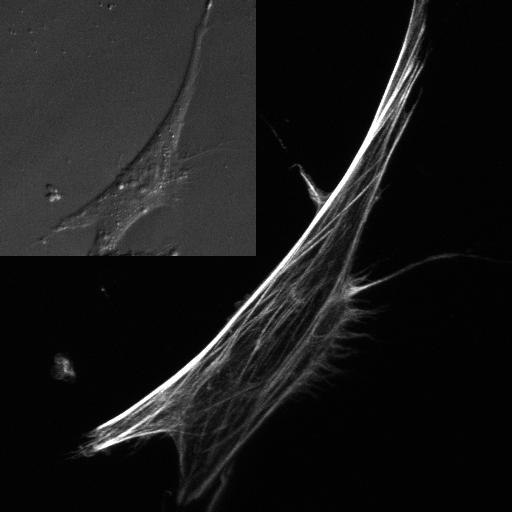
Figure 1.

Fluorescent image of the actin cytoskeleton of a MC3T3-E1 osteoblastic (bone) cell. Microstructurally, the actin cytoskeleton is highly heterogeneous in both the number and orientation of filaments at each point. The inset contains a brightfield image showing the same cell.
