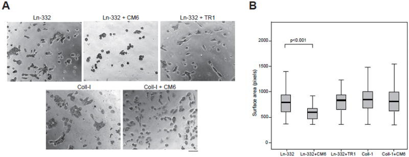
Fig. 5. CM6 inhibits cell spreading on Ln-332.

(A) 96-well microplates were coated with Ln-332 or collagen-I at 4°C overnight. MCF10A cells were labeled with Celltracker™ green CMFDA in PBS (labeling solution) and incubated for 30 min at 37°C. After a washing step, cells were seeded in microplates (5000 cells in 100 μl per well), and allowed to incubate (and spread) for ~7 h, imaging every 6 min using a BD Pathways 855 Bioimager. Normal mouse IgG, CM6, or TR1 were also added where indicated. Representative images from each treatment are shown. Scale bar is equal to 50 μm. (B) Box-and-whisker plots representing mean cell speed (bold horizontal line), 25th and 75th quartiles (box), and 95% confidence intervals (whiskers) are shown (N=70–750). MCF10A cells treated with CM6 exhibited significantly reduced spreading when plated on Ln-332 (p<0.001), but not on collagen-I (p=0.118). In contrast, non-function-blocking antibody TR1 had almost no affect on spreading (p=0.984).
