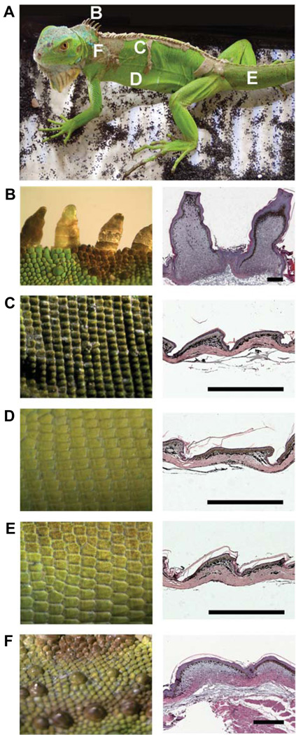
Fig. 3. Arrangement and different types of scales in iguana.
(A) An adult iguana showing different scale types in different regions. (B–F) Left column: scales from regions designated in (A). Right column: H&E staining of their histological sections on the right. (B) Frills from the midline of the neck. Note the elongated scales compared with those in (C–F). (C) Scales from the dorsal trunk. (D) Scales from the ventral trunk. (E) Scales from the tail. (F) Tuberculate scales from the lateral neck region. Scale bars, 500 µm.

