Figure 6.
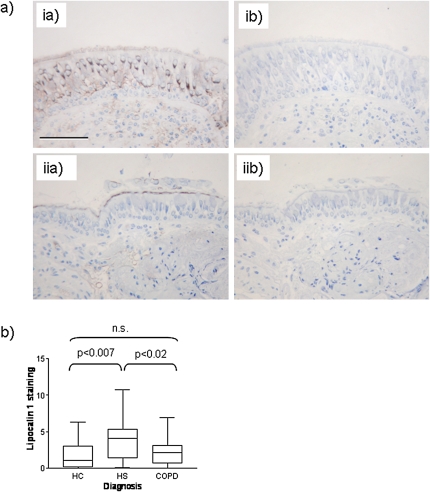
(a) Immunohistochemical staining for lipocalin-1 and apolipoprotein A1 in bronchial tissue. Panel ia: lipocalin-1 staining by specific antibody is observed in the airway epithelium. Panel ib: IgG control for lipocalin. Panel iia: Apolipoprotein staining by specific antibody is observed in the airway lumen and endothelium. Panel iib: IgG control for apolipoprotein A1. In all cases, specific staining is seen in brown; hematoxylin counterstain is in blue. Scale bar: 100 μm. (b) Comparison of staining intensity for lipocalin-1 in the epithelium of nonsmokers, healthy smokers, and smokers with chronic obstructive pulmonary disease (COPD) determined by image analysis and shown as percentage staining of the epithelium. HC = healthy control subjects; HS = healthy smokers; n.s. = not significant.

