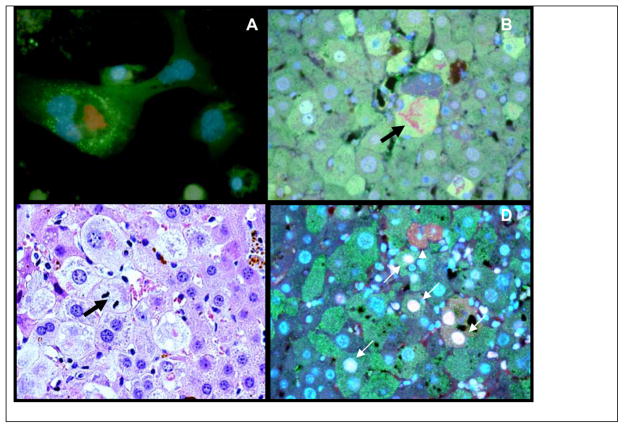
Fig 7.
A) Drug primed tissue culture UB/FAT10. Spontaneous formation of MDB after 6 days in primary culture x800. B) Mouse refed DDC plus SAMe. Note: the red MDB within a Fat10 Positive Cell x800 (Arrow). C) Liver cell in mitosis which had formed a MDB (Arrow) in the liver tissue from a mouse refed DDC 7 Days. Note: The UB positive immunostained MDB forming hepatocytes have a growth advantage (are proliferating) compared to neighboring normal hepatocytes (x800). D) Liver cell from DDC refed mouse double stained with UBD (Green) and PCNA (White) (white arrows). One cell is in telophasis (arrow head) (x800).

