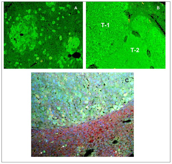
Fig 8.
Immunohistochemical stain of UBD of a mice liver tissue. A) Early liver tumors forming in a background of scattered UBD positive preneoplastic hepatocytes in a mouse withdrawn from DDC 11 months (x200). B) Two Fat10 positive tumors T-1 and T-2 after 8 months DDC withdrawal. C) Mouse liver tumor that stained positive for UBD from a mouse withdrawn from DDC for 8 Months (x80). The tumor cells have formed MDBs. Scaterred single UBD positive hepatocytes are present in the surrounding liver (white arrows).

