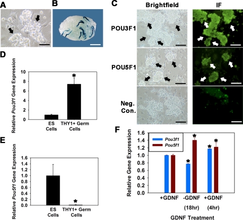
FIG. 1.
Expression of POU3F1 and POU5F1 in cultured mouse THY1+ germ cells. A) Germ cell clumps (arrows) in established cultures of THY1+ germ cells from Rosa donor mouse pups. Bar = 100 μm. B) Recipient mouse testis transplanted with cultured THY1+ germ cells established from Rosa donor mouse pups. Each blue colony of donor spermatogenesis is derived from a single SSC present in the injected cell suspension. Donor spermatogenesis unequivocally demonstrates the presence of SSCs in the cultured cell population. Bar = 2 mm. C) Immunofluorescence analyses of POU3F1 and POU5F1 expression in cultured THY1+ germ cell clumps established from Rosa donor mouse pups. All germ cell clumps (arrows) stain for expression of both molecules. Bars = 100 μm. D and E) Relative Pou3f1 (D) and Pou5f1 (E) gene expression in cultured ES cells and THY1+ germ cells determined using quantitative real-time PCR analysis. Data are the mean ± SEM for three different replicate cultures, and the asterisk denotes significant difference between means. F) Effect of GDNF withdrawal and replacement on expression of Pou3f1 and Pou5f1 in cultured THY1+ germ cells established from Rosa donor mouse pups. Cultures were maintained with GDNF supplementation (+GDNF) and subjected to 18-h withdrawal of the growth factor (−GDNF), followed by replacement for 4 h (+GDNF). Removal of GDNF resulted in down-regulation of Pou3f1 gene expression (blue bars) but up-regulation of Pou5f1 gene expression (red bars). Data are the mean ± SEM for three different replicate cultures, and the asterisk denotes significant difference at P < 0.05 from +GDNF before withdrawal.

