FIG. 2.
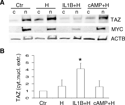
TAZ decline in nucleus during IL1B-induced decidualization. A) Distribution of TAZ in the cytosolic (c) and nuclear (n) fractions of HuF cells treated for 13 days with SHs (H), IL1B and SHs (IL1B+H), cAMP and SHs (cAMP+H), and untreated controls (Ctr) as detected by Western blot with TAZ antibody. Membrane was reprobed with MYC (c-myc) and beta-actin (ACTB) antibodies (blots below). B) Quantification of TAZ distribution in cytosol and nuclear extract fractions of HuF cells after 13 days of decidualization treatments expressed as the cytosol:nuclear extract ratio (labeled as cyt.:nucl. extr.) calculated from densitometric evaluation of TAZ bands from three experiments. The mean ratio in control cells (Ctr) from three experiments was set as 1, and other treatments were compared with the control (mean ± SD). Note the significant increase (*P < 0.05) in the cytosol:nucleus ratio for TAZ in HuF cells treated with IL1B and SHs compared with the control.

