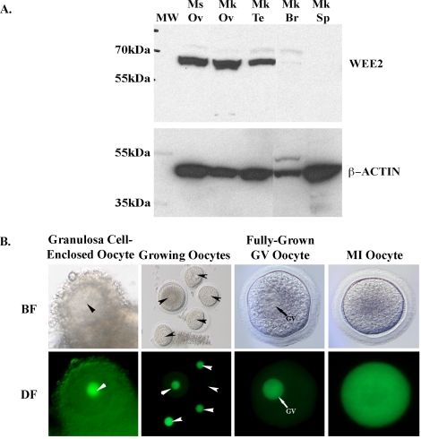
FIG. 2.
Tissue and cellular localization of macaque WEE2 protein. A) Western blot in the mouse ovary and selected monkey tissues. An approximately 65-kDa band was specifically detected in the ovary from both species as well as in the monkey testis. β-ACTIN (42 kDa) was used as an internal loading control. Monkey brain and spleen samples were run on the same gel but not on lanes adjacent to the testis. Br, brain; Mk, monkey; Ms, mouse; MW, molecular weight marker; Ov, ovary; Sp, spleen; Te, testis. B) Distribution of GFP-tagged full-length macaque WEE2 protein in growing oocytes isolated from secondary follicles, fully grown GV, and MI oocytes. Arrowheads denote the nucleus in the oocyte. BF, bright field; DF, dark field. Original magnification ×100 for Growing Oocytes and ×150 for all other panels.

