FIG. 5.
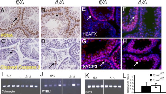
Proliferation, apoptosis, meiosis, and testosterone levels are not altered in Epas1Δ/Δ testes. A and B) Proliferation was not reduced in response to Epas1 deficiency as assessed by IHC for PCNA1 (n = 4) (arrows). C and D) Cleaved caspase-3 was used as a means to visualize apoptotic cells. Epas1Δ/Δ testes did not show increased numbers of dead cells compared with the control testes (arrows). γ-H2AX staining detects double-strand breaks during crossover and remains present in the XY body. E and F) Epas1fl/Δ and Epas1Δ/Δ testes displayed a similar staining pattern (arrows). SCP-3 expression connecting the two sister chromatids was used as a second marker for meiosis. G and H) Testes of both genotypes contained multiple layers of spermatocytes undergoing meiosis (n = 4) (arrows). I–K) The RT-PCR analysis for calmegin (Clgn), Mybl1, and Gpd (used as a loading control) revealed normal expression of these mRNAs. L) Testosterone levels were not changed in Epas1-deficient males. Original magnification ×400 (A–H).

