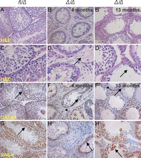
FIG. 6.
The testicular phenotype progresses with age in Epas1Δ/Δ mice. A) Histological analyses of 4-mo-old and 13-mo-old testes obtained from multiple Epas1-deficient males compared with control Epas1fl/Δ testes (A and C). The H&E staining depicted a phenotype worse than that observed at age 2 mo. Epas1Δ/Δ seminiferous tubules had lost increased numbers of germ cells, and the interstitial space was fibrotic (B, B′, D, and D′ [arrows]). GATA4 and VASA expression was used to identify Sertoli cells (E–F′ [arrows]) and germ cells (G–H′ [arrows]); both were still present. GATA4 also detected Leydig cells in the interstitial space (E–F′ [arrowheads]). Original magnification ×200 (A–B′ and E–H′) and ×400 (C–D′).

