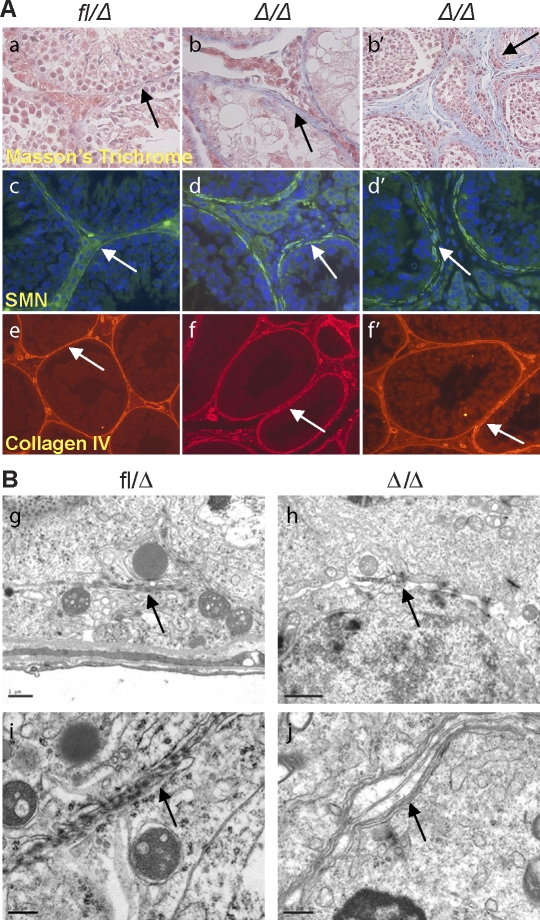
FIG. 7.
EpasΔ/Δ testes exhibit defective basement membrane and myoid cell layers. A) Masson trichrome staining confirmed the fibroblastic character in the interstitial space of Epas1Δ/Δ mice (a, arrow) (original magnification ×400). Epas1Δ/Δ testes aged 4 mo (b) and 13 mo (b′) are shown. Myoid cells were detected by SMA IF (c); control seminiferous tubules were surrounded by a single layer of continuous myoid cells, promoting a tight basement membrane (arrow) (original magnification ×400). However, Epas1Δ/Δ testes displayed thickening and disruption of the basement membrane (d and d′, arrows) (original magnification ×400); the fibroblast-like cells in the interstitial space did not express SMN. Collagen type IV staining delineated irregularity and thickening of the basement membrane of mutant tubules (e–f′, arrows) (original magnification ×200). B) Electron microscopy displayed tight junctions and an impermeable seal between the membranes of two Sertoli cells in control testes (g and i, arrows). Epas1Δ/Δ testes contained shortened tight junctions (arrow in h) and did not form a proper barrier or seal (arrow in j). Furthermore, cells within Epas1Δ/Δ seminiferous tubules were surrounded by multiple membranes (arrow in j). Bar = 1 μm (g, h) and 0.5 μm (i, j).

