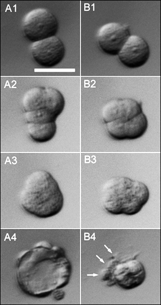
FIG. 1.
Embryo development after splitting two-cell stage embryos into individual blastomeres. Two-cell blastomeres were bisected microsurgically from a single two-cell stage embryo retrieved at 24 h pc and removed from the enclosing ZP. The two sister embryos were cultured individually as demiembryo A (A1–A4) and demiembryo B (B1–B4). At 60 h pc (24 h after bisection), both sisters had cleaved once (A1 and B1). By 72 h pc, the embryos were at the four-cell stage (A2 and B2) and showed signs of early compaction but with different conformations (see also Fig. 3). By 84 h, both embryos (A3 and B3) were around the eight-cell stage and had compacted fully even though they did not have the usual close-to-spherical shape of control morulae. Instead, the contour of the embryos was irregular, and the conformation of individual blastomeres was visible. By 108 h pc, one sister had formed a blastocyst (A4) whereas the other (B4) possessed a compacted cluster of cells and three outer cells (arrowed), one of which possessed a large vacuole. The B4 abnormal blastocyst would be classified as Cav in Tables 1 and 2. Bar = 50 μm.

