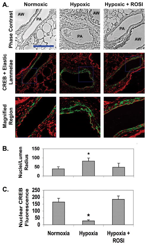
Figure 1. ROSI prevents both PA remodeling and SMC CREB loss in rats exposed to chronic hypoxia.
Sections of lung tissue were prepared from adult male Wistar Kyoto rats were subjected to isobaric normoxia or hypobaric hypoxia for 3 weeks. Some rats exposed to hypoxia were fed chow impregnated with ROSI. At 3 weeks the animals were euthanized, and their lungs were inflated and perfused with PBS containing 4% paraformaldehyde. The sections were deparaffinized, rehydrated and subjected to immunohistochemistry for CREB. A) The top row shows representative phase contrast micrographs of intralobar pulmonary arteries (PA) adjacent to airways (AW). The middle row shows the corresponding fluorescence deconvolution images in which the signal for CREB (red) has been combined with the autofluorescence of the internal and external elastic lamellae (green). The bottom row shows enlargements of the regions within the blue squares in the middle row. Bar = 75 um. B) Cellularity of the medial compartment was quantitated by counting the number of DAPI-stained nuclei located between the inner and outer lammelae. This number was divided by the lumen radius to normalize for vessel size. Data were averaged for at least five vessels per animal from 6 animals. * indicates p ≤ 0.05. C) Nuclear CREB levels were quantitated by measuring fluorescence intensities of nuclei located between the inner and outer lammelae. Data were averaged for at least five vessels per animal from 6 animals. The data indicate that ROSI attenuates hypoxia-induced PA thickening and loss of SMC CREB. * indicates p ≤ 0.05.

