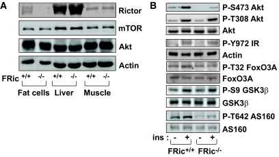
FIG. 1.
Analysis of rictor expression and insulin signaling in FRic−/− fat cells. A: Lack of rictor protein in FRic−/− fat cells. Tissue extracts prepared from isolated fat cells, liver, and skeletal muscle were subjected to SDS-PAGE (6.5% gel) and immunoblotted with anti-rictor antibody (top). The immunoblots for mTOR, Akt, and actin (loading control) are shown in the bottom panels. B: Insulin signaling in isolated FRic−/− fat cells. Immunoblot analysis of insulin (ins)-stimulated phosphorylation of Akt at S473, Akt at T308, IR at Y972, FoxO3A at T32, GSK-3β at S9, and AS160 at T642 in FRic−/− and FRic+/+ fat cells (shown here are representative immunoblots, four mice for each genotype were analyzed). Also shown are immunoblots of total Akt, FoxO3A, GSK-3β, and AS160 used for normalizing the corresponding anti-phospho immunoblots.

