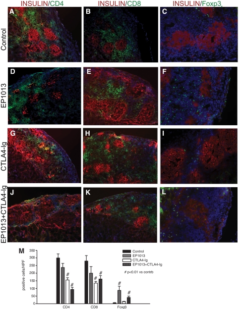
FIG. 4.
Histological analysis in the early post-transplant period demonstrates that EP1013 therapy promotes intragraft accumulation of Foxp3+ lymphocytes. Animals were sacrificed on day 11 post-transplant, and graft-bearing kidneys were harvested for immunfluorescent quantification of intragraft lymphocytic infiltrates. Insulin staining was used to identify transplanted islets (TRITC; red), and the immune markers CD4 (left column), CD8 (middle column), and Foxp3 (right column) were labeled with FITC (green). DAPI counterstaining (blue) was used to identify all cells present within the sections, which are shown at 100× (CD4, CD8) or 200× (Foxp3). Representative sections from each treatment group are shown. M: Quantification of CD4, CD8, and Foxp3 staining was conducted in sections from each treatment group. A significant reduction in the number of CD4+ cells/HPF was observed after treatment with CTLA4-Ig or EP1013+CTLA4-Ig compared with vehicle (P < 0.01 vs. control for both groups). Similar results were observed for CD8+ cells/HPF, with CTLA4-Ig or EP1013+CTLA4-Ig treatment cohorts exhibiting a significant decrease compared with vehicle-treated animals (P < 0.01 vs. control for both groups). Quantification of Foxp3+ cells/HPF revealed that prior treatment with EP1013, either alone or in combination with CTLA4-Ig, resulted in a significant increase in the frequency of intragraft regulatory T-cells (P < 0.01 vs. control and vs. CTLA4-Ig alone by ANOVA). Serial tissue sections from n = 3–4 animals per group were stained, blinded, and analyzed for this experiment. (A high-quality digital color representation of this figure is available in the online issue.)

