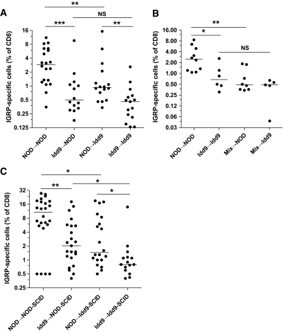
FIG. 6.
Expression of Idd9 genes by both hematopoietic cells and nonlymphocytes contributes to CD8+ T-cell tolerance. Recipient female Thy1.2+ mice were irradiated with 1200 rad and reconstituted with 7 × 10⁶ T-cell–depleted bone marrow cells from Thy1.1+ donors (A) or a 50:50 mix of NOD and Idd9 bone marrow (B). After 12 weeks, the mice were infected with 1 × 10⁷ pfu Vac-IGRP. Splenocytes were stained 7 days later with anti-CD8-FITC and IGRP-tetramer-PE. Pooled data from three experiments are shown. Horizontal line depicts median value. A: NOD→NOD vs. Idd9→NOD P = 0.0006 (***), NOD→NOD vs. NOD→Idd9 P = 0.006 (**), Idd9→NOD vs. Idd9→Idd9 P = 0.24, NOD→Idd9 vs. Idd9→Idd9 P = 0.004 (**), NOD→NOD vs. Idd9→Idd9 P < 0.0001 (Mann-Whitney test). B: NOD→NOD vs. mix→NOD P = 0.005 (**), NOD→NOD vs. Idd9→Idd9 P = 0.016 (Mann-Whitney test). C: NOD-SCID and Idd9-SCID mice were reconstituted with 2 × 10⁷ spleen and LN cells from 3- to 4-week-old NOD or Idd9 mice. After 10 weeks, the mice were infected with Vac-IGRP, and IGRP-tetramer+ CD8+ T-cells were measured in the spleen 7 days later. Pooled data from three experiments are shown.

