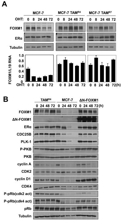
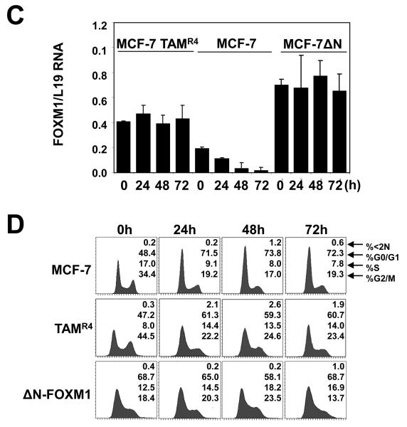
Figure 6. Expression of FOXM1 and cell cycle regulation in wild-type, OHT-resistant, and constitutively active ΔN-FOXM1 expressing MCF-7 cells in response to OHT treatment.
A) MCF-7 and the resistant MCF-7-TAMR4 and -TAMR7 lines cultured in 10% FCS and phenol red medium were treated with OHT in a time course of 72 h. Cell lysates were prepared at the times indicated, and the expression of FOXM1, ERα and tubulin was analyzed by Western blotting. B) MCF-7, MCF-7 TAMR4 and MCF-7 ΔN-FOXM1 cells were treated with OHT in a time course of 72 h. Cell lysates were prepared at the times indicated, and the expression of FOXM1, ERα, CDC25B, P-PKB, total PKB and Tubulin was analyzed by Western blotting. C) FOXM1 mRNA levels of these cells were also analysed by qRT-PCR and normalized to L19 RNA expression. D) Cells were fixed at 0, 24, 48, and 72 h after treatment, and cell cycle phase distribution was analyzed by flow cytometry after propidium iodide staining. Percentage of cells in each phase of the cell cycle (sub-G1, G1, S, and G2/M) is indicated. Representative data from three independent experiments are shown.


