
Fig. 10.
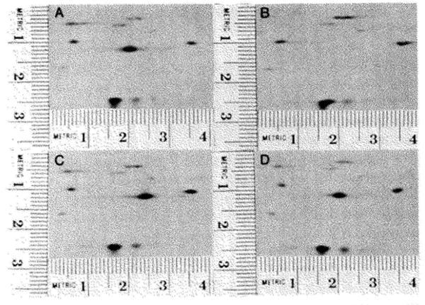
The detection of a missense mutation in T4 gene 32. Samples prepared from T4 infected Escherichia coli (AS19) were subjected to electrophoresis and autoradiograms prepared. Sections of these autoradiograms are shown. The patterns shown were produced by infections with wild type T4 (A) an amber mutant in gene 32 (B), a ts mutant (tsP7) in gene 32 (C), and another ts mutant (tsL170) in gene 32 (D). The spot corresponding to the gene 32 protein was identified by comparing Patterns A and B. A major spot (A, coordinates 2.05 × 1.15) is missing in Pattern B. This spot corresponds to the gene 32 protein. In the pattern shown in Panel C the spot corresponding to the gene 32 protein can still be seen; however, its position (coordinates 2.45 × 1.2) has shifted in comparison to the wild type gene 32. The gene 32 proteins seen in Panel D appears to be in the same position as wild type gene 32 protein. The pattern obtained from the gene 32 amber mutant is missing one minor spot in addition to the gene 32 protein. This minor spot is seen in Panels A, C, and D (coordinates 0.5 × 0.6). It is concluded that this amber mutant (HL618) contains two amber mutations, one in gene 32 and the other in a nonessential gene.