
Fig. 13.
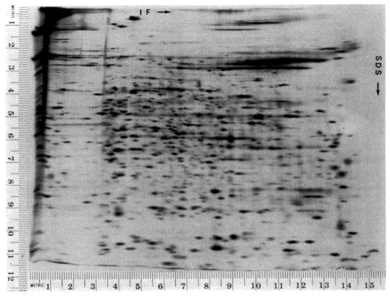
Separation of proteins from Caenorhabdilis elegans. C. elegans was labeled as described under “Materials and Methods,” and lysed by sonication. The lysate was treated with RNase and DNase, and urea and lysis buffer were added. The sample applied to the gel contained 400,000 cpm and 3 μg of protein. The autoradiogram shown was exposed to the gel for 515 hours.