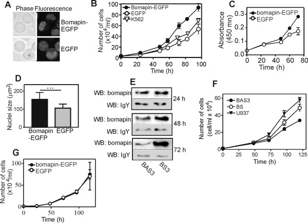
Figure 2.
Wt bomapin promotes proliferation of stably-transfected multi-clonal K562 cells. (A) Cellular localization of bomapin-EGFP and EGFP in transfected K562 cells. (B) Proliferation of the K562 cells expressing bomapin-EGFP and EGFP, and the wt K562 cells (seeded at 2 × 10⁴ cell/ml) measured by manual cell counting. The data represent a mean of three independent experiments, each counted in triplicate. (C) Proliferation of the stably transfected K562 cells measured with the WST-1 reagent. (D) Size of nuclei in K562 cells expressing bomapin-EGFP and EGFP. The symbol "..." indicates statistical significance with p < 0.0001 by unpaired t-test. (E) U937 cells were incubated with bomapin-specific antisense (BAS3) and sense (BS) phosphorothioated DNA oligonucleotides (20 nmol/ml); at different time points bomapin was immunoprecipitated with IgY immobilized on NHS-Sepharose and detected with western blot. Western blot of residual amounts of IgY detached from the beads during immunoprecipitation is shown as loading control. (F) U937 cells were seeded at a density of 1 × 10⁴ cells/ml in the absence or the presence of the antisense BAS3 and corresponding sense BS oligonucleotides, and proliferation was measured by manual counting. The data represent the means of three independent experiments, each counted in triplicate. (G) Proliferation of the HT-1080 cells expressing bomapin-EGFP and EGFP (seeded at 2 × 10⁴ cell/ml) measured by manual cell counting. The data represent a mean of three independent experiments.

