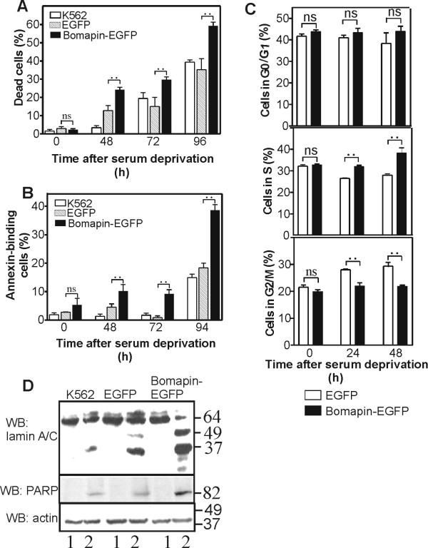
Figure 4.
Bomapin enhances cell apoptosis following growth factors withdrawal. K562 cells and the cells expressing bomapin-EGFP or EGFP were incubated in the presence or the absence of serum in the media. At different time points, cells were mixed with trypan blue and dead cells were quantified by manual counting (A), or the cells were incubated with annexin-PE-Cys5, and annexin-labelled cells were quantified under fluorescence microscope with excitation and emission wavelengths 488 nm and 670 nm, respectively (B); (C) Progression of cell cycle in bomapin-EGFP and EGFP-expressing K562 cells following serum withdrawal. Percentage of G0/G1, S, and G2/M phases were calculated by deconvolution of DNA content histograms; ns - insignificant; ".." indicates statistical significance with p < 0.05. (D) K562 cells expressing bomapin-EGFP or EGFP were incubated in serum-containing media (lanes 1) or in media without serum (lanes 2) for 48 h. Then, cell extracts were analyzed by western blot with monoclonal antibodies against lamins-A/C and rabbit antibodies against cleaved PARP as apoptotic markers. Western blot for β-actin in the same gel is shown as loading control.

