
Figure 3. Bin3 localizes to vesicular membranes that overlap partially with mitochondria but not lysosomes.
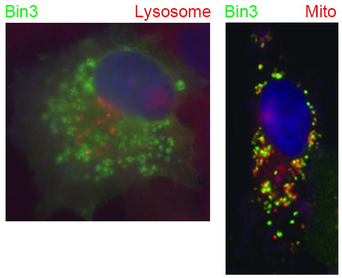
COS cells transfected with a human Bin3 expression vector were fixed and processed for indirect immunofluorescence with a Bin3 antibody [33] and simultaneously stained with DAPI to visualize nuclei plus LysoTracker or MitoTracker, to visualize lysosomes or mitochondria, respectively. Figure is adapted from Ramalingam et al. [34].