
Figure 3.
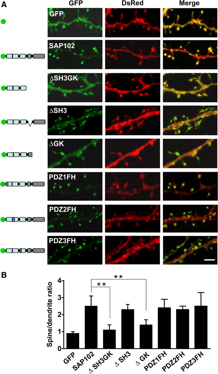
SH3/GK domains are necessary for SAP102 clustering in spines. A, Hippocampal neurons were cotransfected with DsRed/GFP, DsRed/GFP-SAP102, DsRed/GFP-SAP102ΔSH3GK, DsRed/GFP-SAP102ΔSH3, DsRed/GFP-SAP102ΔGK, DsRed/GFP-SAP102PDZ1FH, DsRed/GFP-SAP102PDZ2FH, or DsRed/GFP-SAP102PDZ3FH. Schematic diagrams on the left show the structure of the constructs. GFP-SAP102ΔSH3GK is uniformly distributed throughout the dendrite and the spines. GFP-SAP102ΔSH3, GFP-SAP102PDZ1FH, GFP-SAP102PDZ2FH, and GFP-SAP102PDZ3FH are enriched in spines. Scale bar, 2 μm. B, The mean fluorescence intensity of spines compared with that in adjacent dendrites. The spine/dendrite green fluorescence ratio of GFP-SAP102ΔSH3GK is 1.1 ± 0.3, close to the ratio of the GFP control (0.9 ± 0.1). The ratio of GFP-SAP102ΔSH3, GFP-SAP102PDZ1FH, GFP-SAP102PDZ2FH, and GFP-SAP102PDZ3FH are 2.4 ± 0.3, 2.4 ± 0.5, 2.3 ± 0.2, and 2.5 ± 0.8, all close to the ratio of GFP-SAP102 (2.5 ± 0.6). n = 10–15 neurons from three transfections. **p < 0.01.