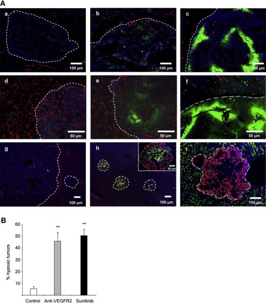
Figure 7. Antiangiogenic Treatment also Provokes Hypoxia in Tumors and Liver Micrometastases.
(A) Hypoxia in islet tumors was detected by immunofluorescence staining of pimonidazole adducts in sections of pancreas (Aa-Af) or liver (Ag-Ai) from control untreated animals (Aa and Ag), animals receiving short-term (Ab) or long-term (Ac) anti-VEGFR2 treatment, β-VEGF-WT (Ad) and β-VEGF-KO (Ae) islet tumors, and sunitinib-treated animals (Af, Ah, and Ai). (Aa)-(Ag) show pimonidazole immunodetection (green) with blood vessel CD31 staining (red); (Ah) shows pimonidazole immunodetection (green) and T antigen onco-protein (red); (Ai) shows blood vessel MECA32 staining (green) and T antigen oncoprotein (red). All images show nuclei counterstained with DAPI (blue).
(B) Quantitation of the incidence of hypoxic tumors was performed in long-term anti-VEGFR2-treated and sunitinib-treated animals and plotted as the percentage of pimonidazole-positive tumors per animal compared to control animals. **p < 0.01 by Mann-Whitney test. Error bars indicate ± SEM.

