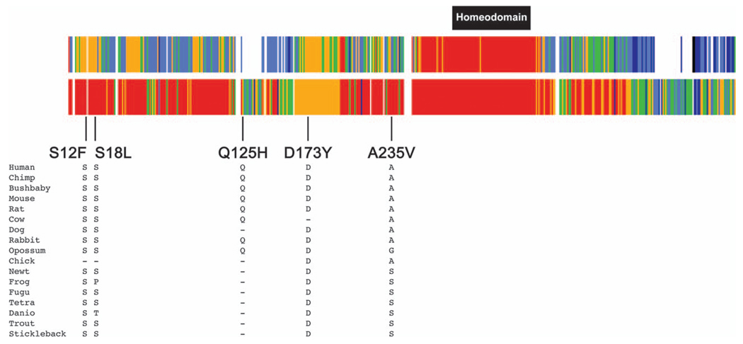
Fig. 2.
Alignment of the HLX gene showing conserved residues at the site of the sequence variants predicting p.S12F, p.S18L, p.D173Y and p.A235V. The top bar shows conservation among the 17 species shown in the figure. Red indicates that the amino acids are identical in all 17 species; orange indicates that the amino acids are identical in 14–16 species; green indicates that the amino acids are identical in 11–12 species; light blue indicates that the amino acids are identical in 7–10 species; dark blue indicates that the amino acids are identical in four to six species; black indicates that the amino acids are identical in three species and white indicates that the amino acids are identical in one to two species. The second bar shows conservation among the eight mammalian species. Red indicates that the amino acids are identical in all eight species; orange indicates that the amino acids are identical in seven species; green indicates that the amino acids are identical in five to six species; light blue indicates that the amino acids are identical in four species; dark blue indicates that the amino acids are identical in three species.

