
Figure 6.
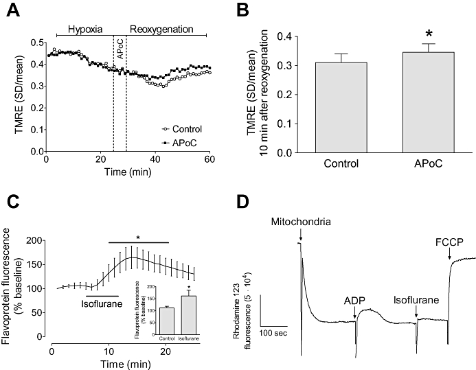
APoC preserves ΔΨm in isolated myocytes throughout reoxygenation. (A) Representative recording of ΔΨm changes during H/R. Hypoxia caused a significant decrease in the standard deviation of TMRE fluorescence indicating mitochondrial depolarization whereas reoxygenation caused an increase in the standard deviation indicating recovery of ΔΨm. Treatment of cardiomyocytes with isoflurane at the beginning of reoxygenation preserved ΔΨm as compared with control cells. (B) Summary data for control and APoC groups at 10 min of reoxygenation. (C) Effect of isoflurane on mitochondrial flavoprotein fluorescence, and summary data (inset). (D) Representative trace of ΔΨm in isolated mitochondria. Isoflurane induced slight depolarization of ΔΨm. Data are means ± standard deviation; n= 15; n= 6 in flavoprotein and isolated mitochondria experiments. *P < 0.05 versus control. APoC, isoflurane postconditioning, TMRE, tetramethylrhodamine ethyl ester. FCCP, carbonyl cyanide 4-(trifluoromethoxy)phenylhydrazone.