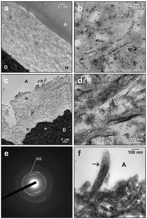
Figure 2.
Transmission electron micrographs showing examples of intrafibrillar remineralization in resin-dentin interfaces after 2-3 mos of biomimetic remineralization. C, composite; A, unfilled adhesive; FA, filled adhesive; H, hybrid layer; D, mineralized dentin. (a) A One-Step specimen taken after 2 mos, with remineralization (pointer) observable at low magnification. (b) High magnification of (a), showing nanocrystals along the interfibrillar spaces (between open arrows). The earliest stage of intrafibrillar remineralization could be recognized as an ordered alignment of nanocrystals (open arrowheads). (c) A One-Step specimen taken after 3 mos, with remineralization occurring in the middle of the hybrid layer and along the surface collagen fibrils (arrow). Regions that were not remineralized (asterisks) were probably better infiltrated with adhesive resin. (d) A high-magnification view of the region depicted by the “pointer” in (c), illustrating a more advanced stage of intrafibrillar remineralization. Crystallite platelets (ca. 20 nm long) were stacked in a repeating and orderly sequence (triple open arrowheads) within the collagen fibrils (ca. 100 nm in diameter). The crystallographic c-axes of these electron-dense platelets were well-aligned with the axis of the collagen fibrils. Larger needle-shaped crystallites (ca. 50 nm) could be seen (pointers) along the periphery of the fibrils (i.e., interfibrillar remineralization). (e) SAED of the region in (d). The arc-shaped diffraction patterns ascribed to the (002) plane of apatite suggest that the c-axes of the platelets have a preferential orientation. (f) A high-magnification view of (c), showing the density of apatite platelets within a remineralized collagen fibril (arrow) along the hybrid layer surface.

