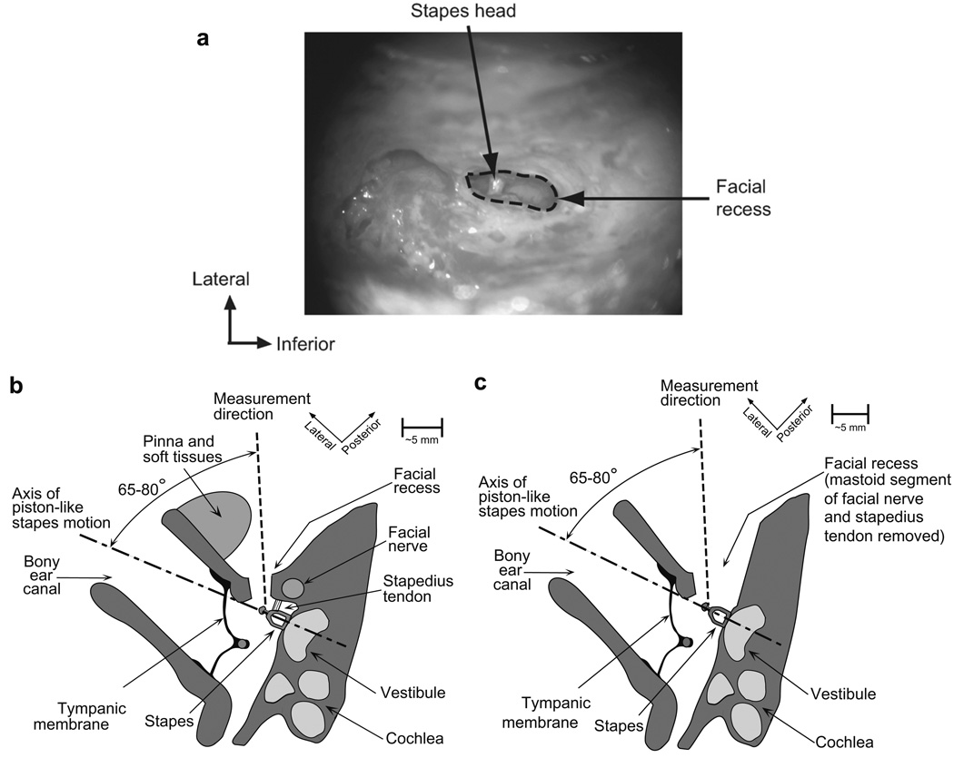
Fig. 3.
(A) Photograph of an intraoperative view of the stapes during Vs measurement (right ear). The stapes head is shown with a naturally reflective spot where Vs measurements were made. (B) Schematic axial view of the measurement setup in live Vs measurements. (C) Schematic axial view of the measurement setup in cadaveric Vs measurements. Note that even though the measurement direction is aimed at the head of stapes, some of our cadaveric Vs measurements were made at the posterior crus, as indicated in the text.

