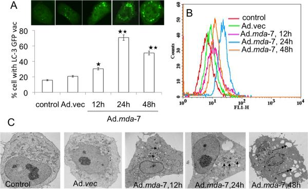
Fig. 1. MDA-7/IL-24 induces autophagy in DU-145 cells.
A, DU-145 cells were transfected with GFP-LC3 and infected with Ad.mda-7 (100pfu/cell) and after different times localization of LC3 in transfected cells was examined by confocal microscopy (magnification ×100) and autophagosome formation was quantified and data presented as percentage of GFP-LC3–transfected cells with punctate fluorescence to autophagosome formation. A minimum of 100 GFP-LC3–transfected cells were counted. *, P < 0.05; **, P < 0.001, compared with control at the corresponding time. B, The MDC fluorescent intensity of Ad.mda-7-treated DU-145 cells was analyzed by flow cytometry. This result was representative of three different experiments. C, DU-145 cells were infected with 100 pfu/cell of Ad.mda-7 or Ad.vec for different times and cells were fixed and processed for electron microscopy (single arrow, autophagosome; double arrow, empty vacuole; Scale bars: control and others-2μm).

