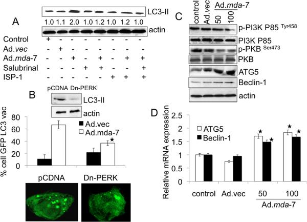
Fig. 3. MDA-7/IL-24-induced autophagy is mediated through ER stress and ceramide production.
A, DU-145 cells were infected with 100 pfu/cell of Ad.mda-7 or Ad.vec in the presence of Salubrinal (5 μM) and/or ISP-1 (10 μM) for 24 h followed by analysis of LC3 expression by immunoblotting. Densitometry was performed on the original blots, and the ratio of LC3-II/actin in control cells was 1. B, DU-145 cells were transfected with the indicated plasmids: empty vector control plasmid (pCDNA) or a plasmid expressing dominant negative PERK (Dn-PERK) and GFP-LC3. Cells were analyzed for LC3 aggregation and LC3 expression 24 h after Ad.mda-7 or Ad.vec infection (100 pfu/cell). *, P < 0.05; compared with pCDNA Ad.mda-7 infected cells. C, Evaluation of protein expression: Beclin-1, ATG5, total PKB, p-PKB, total PI3K and p-PI3K using immunoblotting 24 h after Ad.mda-7 or Ad.vec infection (100 pfu/cell). D, DU-145 cells were infected with 100 pfu/cell of Ad.mda-7 or Ad.vec and after 24 h expression of Beclin-1 and ATG5 mRNA was determined using Taqman real-time PCR. Values are the mean ± S.D. of three independent experiments and *, p < 0.05 versus control cells.

